
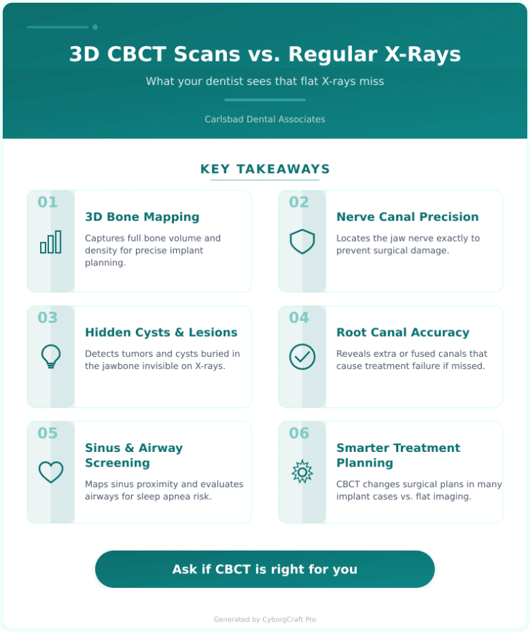
A 3D CBCT scan (Cone Beam Computed Tomography) is a focused dental imaging technique that captures a full three-dimensional picture of your teeth, jaw, nerves, sinuses, and bone structure in a single low-dose rotation. Unlike a flat X-ray, it gives your dentist a complete volumetric view that reveals hidden problems before they become serious.
This guide focuses specifically on what CBCT imaging detects, why it matters for treatment planning, and what Carlsbad patients should expect during and after the scan.
CBCT Imaging Definition: Cone Beam Computed Tomography is a diagnostic imaging technology used in dentistry to produce three-dimensional images of bone, teeth, nerve pathways, and soft tissue in one low-radiation scan.
If your dentist recommended a CBCT scan, your first instinct might be to wonder whether it was really necessary. Here’s the honest answer: for certain conditions, a traditional two-dimensional X-ray simply cannot show what needs to be seen. In some clinical situations, relying only on two-dimensional imaging can miss information that three-dimensional imaging provides.
What a Standard Dental X-Ray Actually Shows
Conventional dental X-rays, including bitewings and periapical films, are flat images. They’re excellent for spotting cavities between teeth, checking bone levels around existing teeth, and monitoring fillings. For routine checkups, they do the job well.
But here’s the limitation: a two-dimensional image collapses a three-dimensional structure. Overlapping anatomy hides detail. A root that curves away from the sensor, a cyst tucked behind the jawbone, or a nerve canal that splits unexpectedly – none of these show up clearly on a standard film.
According to the American Dental Association, CBCT imaging is recommended when two-dimensional imaging is insufficient for clinical decision-making – particularly for implant planning, complex extractions, and evaluating jaw pathology.
What a 3D CBCT Scan Catches That X-Rays Miss
The difference between flat imaging and CBCT is significant for specific clinical situations. Here are the most common findings that only show up in three dimensions:
- Bone volume and density at implant sites – critical for determining whether enough structure exists to support an implant
- Precise nerve canal location – the inferior alveolar nerve runs through the lower jaw, and nicking it during surgery causes numbness or pain
- Hidden cysts, tumors, or lesions buried in the jawbone
- Root fractures that don’t appear on standard films
- Extra or fused root canals that would cause treatment failure if missed
- Sinus proximity when placing upper back implants
- Airway evaluation for sleep apnea screening
- Impacted wisdom teeth and their exact relationship to adjacent roots
Recent data shows that CBCT imaging changes the surgical treatment plan in a meaningful percentage of implant cases compared to planning from conventional X-rays alone. That matters because a plan built on incomplete information leads to complications that are far more difficult and expensive to fix.
3D CBCT vs Traditional X-Rays: Which Approach Works?
Where traditional X-rays succeed: Routine cavity detection, monitoring bone levels in recall appointments, checking restoration fit, low cost, low radiation dose for simple diagnostic needs.
Where traditional X-rays fail: Cannot show depth or volume, miss hidden jaw pathology, inadequate for implant planning or complex surgical cases, overlapping anatomy creates blind spots.
Where CBCT imaging succeeds: Full three-dimensional bone mapping, precise nerve and sinus localization, detects cysts and lesions invisible on flat films, supports accurate implant and surgical planning.
Where CBCT imaging fails: Higher radiation dose than a standard X-ray (though still significantly lower than a medical CT scan), higher cost, not necessary for every patient or appointment.
The verdict: CBCT is not a replacement for routine X-rays – it’s the right tool for specific, higher-stakes diagnostic situations. If your dentist ordered one, it means your case has a clinical complexity that flat imaging cannot safely address.
| Feature | Standard X-Ray | 3D CBCT Scan |
|---|---|---|
| Image Type | 2D flat film | 3D volumetric |
| Typical Cost | Varies by practice and location | Varies by practice and location |
| Radiation Dose | Very low | Low (less than medical CT) |
| Best For | Routine exams, cavities | Implants, surgery, pathology |
| Nerve Mapping | No | Yes |
| Bone Volume Measurement | No | Yes |
Thinking about what your scan results mean for your treatment? Contact us and we’ll walk you through your imaging findings clearly – no pressure, no jargon.
What to Expect During a CBCT Scan
Phase 1: Preparation (5 minutes)
You’ll remove any metal objects including jewelry, glasses, and removable dental appliances. A lead apron may be placed over your chest and abdomen as a precaution.
Key actions during this phase:
- Remove earrings, necklaces, and hair clips
- Let the assistant know if you are pregnant
Phase 2: Imaging (20-40 seconds)
You’ll stand or sit while the CBCT unit rotates around your head in a single pass. You’ll need to stay still. The machine does not touch you. Most patients describe it as completely painless and far less uncomfortable than a traditional mold or impression.
Phase 3: Review (Same day or next visit)
Your dentist reviews the three-dimensional model on-screen, measures bone dimensions, maps nerve pathways, and identifies any unexpected findings before confirming or adjusting the treatment plan.
Your CBCT Scan Action Plan
- Step 1 – Ask why: Before the scan, ask your dentist specifically what they are looking for. Understanding the purpose helps you engage with results later.
- Step 2 – Disclose your health history: Mention pregnancy, recent radiation exposure, or implanted devices so your provider can adjust protocols appropriately.
- Step 3 – Review images together: Ask your dentist to walk through the three-dimensional images with you. Most modern software lets you see the scan on a monitor during the consultation.
- Step 4 – Confirm the treatment plan change: Ask whether the CBCT findings changed the original plan and how.
- Step 5 – Verify insurance coverage: CBCT scans are covered by some dental plans when medically necessary. Ask the front desk to check your benefits before the appointment.
Key Takeaways for Carlsbad Patients in 2026
- CBCT imaging fills a real diagnostic gap – flat X-rays cannot show bone depth, nerve location, or hidden jaw pathology with the accuracy needed for surgical cases.
- The radiation dose is low – significantly less than a medical CT scan and comparable to a few standard dental X-rays depending on field size.
- It directly affects your treatment outcome – implant placement, extractions, and root canal planning all improve when built on three-dimensional data.
- Costs vary by practice, geography, and insurance coverage – insurance often covers the scan when deemed medically necessary for a specific procedure.
- You should always ask to review the images – seeing your own scan in 3D is both informative and reassuring.
At Carlsbad Dental Associates, our team uses CBCT imaging as a precision diagnostic tool when clinical complexity demands it – not as a routine upsell. Patients from Carlsbad, Oceanside, Vista, San Marcos, Encinitas, and the broader North County San Diego area visit our office at 5814 Van Allen Way #220, Carlsbad, CA 92008. For a complete overview of our diagnostic and treatment services, visit our services page.
The National Institute of Dental and Craniofacial Research supports the use of advanced imaging technologies where they improve diagnostic accuracy and patient outcomes – and the clinical evidence for CBCT in implant and surgical planning is well established.
Disclaimer: This content is for educational purposes only and is not a substitute for professional dental advice, diagnosis, or treatment. Always consult a licensed dental professional regarding your specific oral health needs.
Frequently Asked Questions
Is a 3D CBCT scan safe, and how much radiation does it involve?
Yes, CBCT scans are considered safe and use significantly less radiation than a hospital CT scan. Depending on the field of view selected, the dose is often comparable to a small series of conventional dental X-rays. Your dentist follows the ALARA principle (as low as reasonably achievable) when prescribing any imaging.
How much does a CBCT scan cost without insurance?
Out-of-pocket costs for a CBCT scan vary depending on the field of view, the practice, and your location. Larger full-arch scans cost more than focused single-arch images. Many dental insurance plans cover the scan when it is required for a specific procedure like implant placement.
Does a CBCT scan hurt?
No – a CBCT scan is completely painless. The machine rotates around your head without any contact. You simply hold still for roughly 20 to 40 seconds while the unit captures the image.
Why did my dentist order a CBCT scan before placing an implant?
Implant placement requires precise knowledge of bone volume, bone density, and nerve location – none of which a flat X-ray can provide reliably. Without three-dimensional imaging, a dentist cannot safely confirm that enough bone exists or that the planned implant position avoids the inferior alveolar nerve.
Can a CBCT scan detect oral cancer or jaw tumors?
CBCT imaging can reveal suspicious lesions, cysts, and abnormal bone changes that may require further investigation. It is not a definitive cancer diagnostic tool on its own, but it often flags irregularities that prompt biopsy or referral to an oral surgeon for closer evaluation.
How often should a patient get a CBCT scan?
CBCT scans are not routine – they are prescribed for specific clinical indications. Your dentist will recommend one only when the diagnostic benefit outweighs the radiation exposure, such as before surgery, implant placement, or evaluation of an identified jaw abnormality.